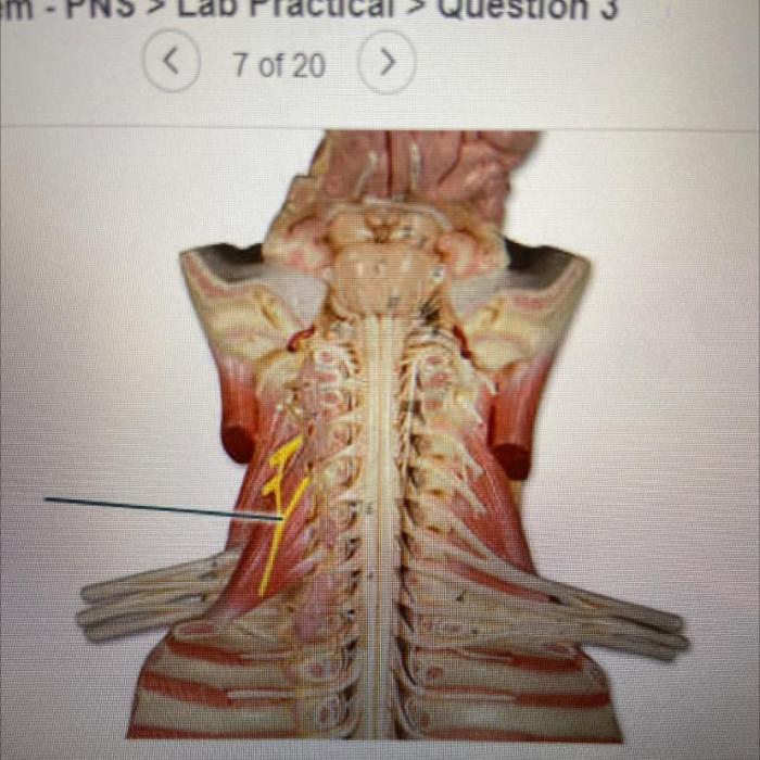
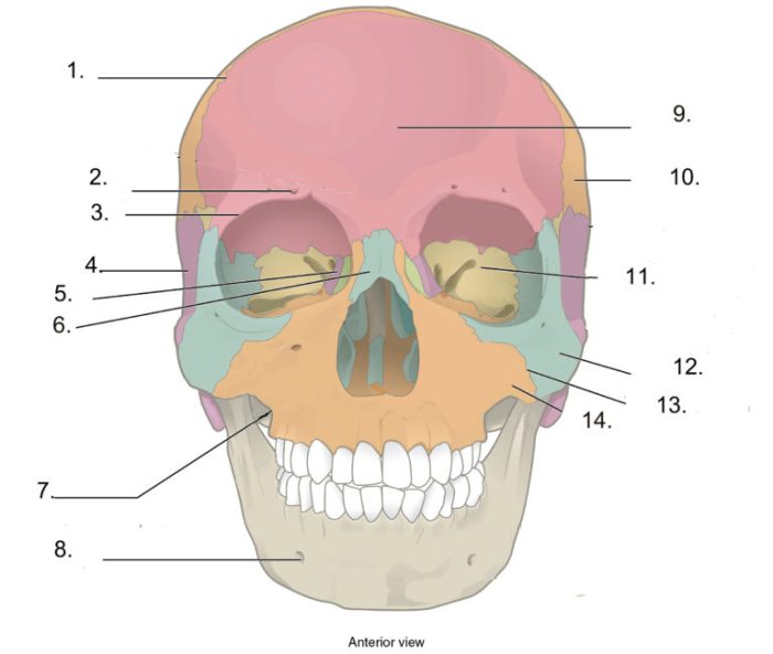
Identify the highlighted structure bone – Embarking on a journey of anatomical discovery, we delve into the intricacies of the highlighted structure bone, a pivotal component of our skeletal system. This bone, meticulously crafted by nature, plays a multifaceted role in our movements, postures, and overall skeletal stability.
Its significance extends beyond its structural contributions, as it serves as a valuable diagnostic tool in various musculoskeletal disorders. Join us as we unravel the secrets of this enigmatic bone, examining its anatomical landmarks, embryological development, biomechanical properties, and clinical implications.
Overview of the Humerus

The humerus is the longest bone in the upper limb, extending from the shoulder joint to the elbow joint. It consists of a shaft and two ends, the proximal end and the distal end. The proximal end of the humerus articulates with the glenoid cavity of the scapula, forming the shoulder joint.
The distal end of the humerus articulates with the radius and ulna, forming the elbow joint.The humerus is responsible for various movements of the upper limb, including flexion, extension, abduction, adduction, and rotation. It also provides structural support to the upper limb and protects the neurovascular structures within.
Anatomical Landmarks of the Humerus
Proximal End
- Greater tubercle: Site of attachment for the supraspinatus muscle.
- Lesser tubercle: Site of attachment for the subscapularis muscle.
- Surgical neck: Region where the shaft of the humerus meets the proximal end.
Shaft
- Deltoid tuberosity: Site of attachment for the deltoid muscle.
- Radial groove: Groove that accommodates the radial nerve.
Distal End, Identify the highlighted structure bone
- Capitulum: Articulates with the radius.
- Trochlea: Articulates with the ulna.
- Medial and lateral epicondyles: Sites of attachment for various muscles and ligaments.
The anatomical landmarks of the humerus are crucial for surgical procedures and clinical examinations. Variations in these landmarks can affect the biomechanics of the upper limb and may be associated with certain pathological conditions.
Embryological Development of the Humerus

The humerus develops from a cartilaginous model that appears during the fifth week of gestation. The ossification of the humerus begins at the proximal end and progresses towards the distal end. The proximal epiphysis fuses with the shaft at around 18 years of age, while the distal epiphysis fuses at around 16 years of age.The
development of the humerus is regulated by a complex interplay of molecular and genetic factors. Mutations in certain genes, such as GLI3 and PTHLH, have been linked to congenital anomalies of the humerus, such as brachydactyly and polydactyly.
Role of the Humerus in Movements and Postures
The humerus plays a crucial role in various movements of the upper limb, including:
- Flexion: Bending of the elbow joint.
- Extension: Straightening of the elbow joint.
- Abduction: Raising the arm away from the body.
- Adduction: Lowering the arm towards the body.
- Rotation: Turning the palm up (supination) or down (pronation).
The humerus provides structural support to the upper limb and protects the neurovascular structures within. The muscles that attach to the humerus, such as the biceps brachii and triceps brachii, generate the forces necessary for these movements.
Clinical Significance of the Humerus
The humerus is of great clinical significance due to its involvement in various musculoskeletal disorders. Fractures of the humerus are common, particularly in the proximal and distal ends. These fractures can occur due to trauma, falls, or sports injuries.Imaging techniques such as X-rays and CT scans are used to diagnose fractures and assess the extent of the injury.
Treatment options for humeral fractures vary depending on the severity and location of the fracture. Conservative treatment with immobilization may be sufficient for some fractures, while others may require surgical intervention.The humerus is also affected by various pathological conditions, such as osteoarthritis and rheumatoid arthritis.
These conditions can cause pain, stiffness, and swelling of the shoulder and elbow joints. Treatment for these conditions may involve medication, physical therapy, or surgery.
Comparison of the Humerus with Other Bones: Identify The Highlighted Structure Bone
The humerus can be compared to other long bones in the body, such as the femur and tibia. Similarities include their overall structure, consisting of a shaft and two ends. They also share similar functions, such as providing structural support and facilitating movement.However,
there are also some key differences between the humerus and other long bones. The humerus is unique in its role in the complex movements of the upper limb, including rotation. Additionally, the anatomical landmarks of the humerus, such as the greater and lesser tubercles, are distinctive and play important roles in muscle attachments and joint stability.
Detailed FAQs
What is the primary function of the highlighted structure bone?
The highlighted structure bone plays a crucial role in supporting and protecting vital organs, facilitating movement, and providing leverage for muscle attachment.
How can variations in anatomical landmarks affect surgical procedures?
Variations in anatomical landmarks can impact the accuracy and safety of surgical interventions, necessitating careful preoperative planning and intraoperative adjustments.
What are some congenital anomalies associated with the development of the highlighted structure bone?
Congenital anomalies, such as aplasia (absence of bone formation) or hypoplasia (underdevelopment), can occur during the embryological development of the highlighted structure bone.